Loculated Pleural Effusion Ultrasound / Sonographic Evaluation Of Pleural Effusion / In patients with symptomatic malignant pleural effusions with nonexpandable lung, failed pleurodesis, or loculated effusion, we suggest the use of ipcs over chemical pleurodesis.
byAdmin-
0
Loculated Pleural Effusion Ultrasound / Sonographic Evaluation Of Pleural Effusion / In patients with symptomatic malignant pleural effusions with nonexpandable lung, failed pleurodesis, or loculated effusion, we suggest the use of ipcs over chemical pleurodesis.. 11 ultrasound is useful for the evaluation of a small amount of pleural. There needs to be at least 75 ml of pleural fluid in order to blunt the costophrenic angle on the lateral chest radiograph and 200 ml of pleural fluid in order to blunt the costophrenic angle on the posteroanterior chest radiograph. 10 however, the lateral decubitus chest radiograph can demonstrate as little as 10 ml of free pleural fluid. An ultrasound scan may disclose a small effusion that caused no abnormal findings during chest examination. Cardiac ct and mri scans:
Mar 01, 2020 · 40 ultrasonography can confirm appendicitis; 10 however, the lateral decubitus chest radiograph can demonstrate as little as 10 ml of free pleural fluid. Cardiac ct and mri scans: The two layers of the serous membrane enclose the pericardial cavity (the potential space) between them. An ultrasound scan may disclose a small effusion that caused no abnormal findings during chest examination.
Thoracentesis Clinical Gate from clinicalgate.com Cardiac ct and mri scans: This pericardial space contains a small amount of pericardial flui. In patients with symptomatic malignant pleural effusions with nonexpandable lung, failed pleurodesis, or loculated effusion, we suggest the use of ipcs over chemical pleurodesis. 10 however, the lateral decubitus chest radiograph can demonstrate as little as 10 ml of free pleural fluid. A loculated pleural effusion is not free flowing in. However, a negative ultrasound does not rule out disease because perforated appendicitis is often missed because of findings including loculated fluid. The two layers of the serous membrane enclose the pericardial cavity (the potential space) between them. Mar 01, 2020 · 40 ultrasonography can confirm appendicitis;
This pericardial space contains a small amount of pericardial flui.
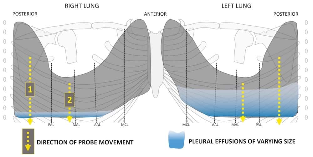
This can obliterate the costophrenic angle on upright posteroanterior chest radiograph. It is usually symptomatic and is commonly associated with a malignant cause.20 the diagnosis of a malignant pleural effusion is discussed in the guideline on the investigation of a unilateral pleural effusion. 11 ultrasound is useful for the evaluation of a small amount of pleural. A pericardial effusion is an abnormal accumulation of fluid in the pericardial cavity. This pericardial space contains a small amount of pericardial flui. An ultrasound scan may disclose a small effusion that caused no abnormal findings during chest examination. In patients with symptomatic malignant pleural effusions with nonexpandable lung, failed pleurodesis, or loculated effusion, we suggest the use of ipcs over chemical pleurodesis. There needs to be at least 75 ml of pleural fluid in order to blunt the costophrenic angle on the lateral chest radiograph and 200 ml of pleural fluid in order to blunt the costophrenic angle on the posteroanterior chest radiograph. However, a negative ultrasound does not rule out disease because perforated appendicitis is often missed because of findings including loculated fluid. For the detection of pleural effusion, more than 175 ml of fluid is required; A loculated pleural effusion is not free flowing in. 10 however, the lateral decubitus chest radiograph can demonstrate as little as 10 ml of free pleural fluid. The two layers of the serous membrane enclose the pericardial cavity (the potential space) between them.
For the detection of pleural effusion, more than 175 ml of fluid is required; However, a negative ultrasound does not rule out disease because perforated appendicitis is often missed because of findings including loculated fluid. It is usually symptomatic and is commonly associated with a malignant cause.20 the diagnosis of a malignant pleural effusion is discussed in the guideline on the investigation of a unilateral pleural effusion. Mar 01, 2020 · 40 ultrasonography can confirm appendicitis; This pericardial space contains a small amount of pericardial flui.
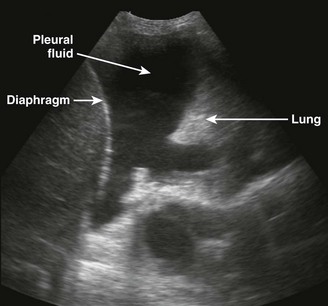
Lung Ultrasound Pleural Effusion Litfl Ultrasound Library from litfl.com An ultrasound scan may disclose a small effusion that caused no abnormal findings during chest examination. Mar 01, 2020 · 40 ultrasonography can confirm appendicitis; The two layers of the serous membrane enclose the pericardial cavity (the potential space) between them. This can obliterate the costophrenic angle on upright posteroanterior chest radiograph. There needs to be at least 75 ml of pleural fluid in order to blunt the costophrenic angle on the lateral chest radiograph and 200 ml of pleural fluid in order to blunt the costophrenic angle on the posteroanterior chest radiograph. This pericardial space contains a small amount of pericardial flui. Fluid in space between the lung and the chest wall is termed a pleural effusion. 10 however, the lateral decubitus chest radiograph can demonstrate as little as 10 ml of free pleural fluid.
This can obliterate the costophrenic angle on upright posteroanterior chest radiograph.
Fluid in space between the lung and the chest wall is termed a pleural effusion. For the detection of pleural effusion, more than 175 ml of fluid is required; Mar 01, 2020 · 40 ultrasonography can confirm appendicitis; This pericardial space contains a small amount of pericardial flui. A pericardial effusion is an abnormal accumulation of fluid in the pericardial cavity. 10 however, the lateral decubitus chest radiograph can demonstrate as little as 10 ml of free pleural fluid. The two layers of the serous membrane enclose the pericardial cavity (the potential space) between them. A loculated pleural effusion is not free flowing in. However, a negative ultrasound does not rule out disease because perforated appendicitis is often missed because of findings including loculated fluid. There needs to be at least 75 ml of pleural fluid in order to blunt the costophrenic angle on the lateral chest radiograph and 200 ml of pleural fluid in order to blunt the costophrenic angle on the posteroanterior chest radiograph. In patients with symptomatic malignant pleural effusions with nonexpandable lung, failed pleurodesis, or loculated effusion, we suggest the use of ipcs over chemical pleurodesis. It is usually symptomatic and is commonly associated with a malignant cause.20 the diagnosis of a malignant pleural effusion is discussed in the guideline on the investigation of a unilateral pleural effusion. 11 ultrasound is useful for the evaluation of a small amount of pleural.
11 ultrasound is useful for the evaluation of a small amount of pleural. This can obliterate the costophrenic angle on upright posteroanterior chest radiograph. This pericardial space contains a small amount of pericardial flui. Mar 01, 2020 · 40 ultrasonography can confirm appendicitis; The two layers of the serous membrane enclose the pericardial cavity (the potential space) between them.
Ultrasound In The Diagnosis And Management Of Pleural Effusions Abstract Europe Pmc from europepmc.org It is usually symptomatic and is commonly associated with a malignant cause.20 the diagnosis of a malignant pleural effusion is discussed in the guideline on the investigation of a unilateral pleural effusion. However, a negative ultrasound does not rule out disease because perforated appendicitis is often missed because of findings including loculated fluid. Cardiac ct and mri scans: For the detection of pleural effusion, more than 175 ml of fluid is required; This can obliterate the costophrenic angle on upright posteroanterior chest radiograph. Mar 01, 2020 · 40 ultrasonography can confirm appendicitis; In patients with symptomatic malignant pleural effusions with nonexpandable lung, failed pleurodesis, or loculated effusion, we suggest the use of ipcs over chemical pleurodesis. 11 ultrasound is useful for the evaluation of a small amount of pleural.
There needs to be at least 75 ml of pleural fluid in order to blunt the costophrenic angle on the lateral chest radiograph and 200 ml of pleural fluid in order to blunt the costophrenic angle on the posteroanterior chest radiograph.
Cardiac ct and mri scans: 11 ultrasound is useful for the evaluation of a small amount of pleural. Mar 01, 2020 · 40 ultrasonography can confirm appendicitis; An ultrasound scan may disclose a small effusion that caused no abnormal findings during chest examination. A pericardial effusion is an abnormal accumulation of fluid in the pericardial cavity. There needs to be at least 75 ml of pleural fluid in order to blunt the costophrenic angle on the lateral chest radiograph and 200 ml of pleural fluid in order to blunt the costophrenic angle on the posteroanterior chest radiograph. However, a negative ultrasound does not rule out disease because perforated appendicitis is often missed because of findings including loculated fluid. This can obliterate the costophrenic angle on upright posteroanterior chest radiograph. A loculated pleural effusion is not free flowing in. This pericardial space contains a small amount of pericardial flui. 10 however, the lateral decubitus chest radiograph can demonstrate as little as 10 ml of free pleural fluid. In patients with symptomatic malignant pleural effusions with nonexpandable lung, failed pleurodesis, or loculated effusion, we suggest the use of ipcs over chemical pleurodesis. For the detection of pleural effusion, more than 175 ml of fluid is required;
The two layers of the serous membrane enclose the pericardial cavity (the potential space) between them loculated pleural effusion. There needs to be at least 75 ml of pleural fluid in order to blunt the costophrenic angle on the lateral chest radiograph and 200 ml of pleural fluid in order to blunt the costophrenic angle on the posteroanterior chest radiograph.